
Image Cytometry
Image Cytometry - Instruments
-
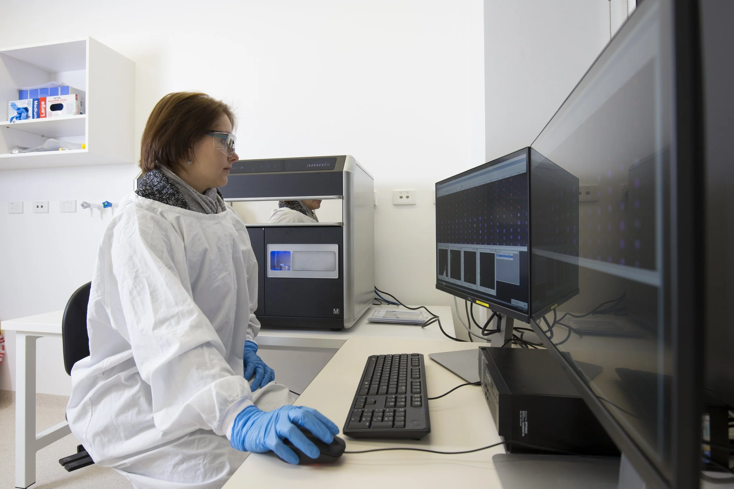
Amnis ImageStream MKII
Overview: 5 Laser, 12 parameter flow cytometer that takes an image of each cell, across each channel. Providing both locational and standard flow cytometer scatter information
Lasers: Violet, Blue, Yellow-Green, Red and IR (for scatter)
9 colour + 1 scatter + 2 brightfield parametersSample acquisition of up to 5000 events/second
Capabilities:
- 3 Magnifications - 20x, 40x, 60x
- 96-well autosampler, plus standard tube acquisition
- Dedicated analysis software -

PerkinElmer Opera Phenix Plus
Overview: 5 Laser, 4 Camera, Confocal High-Content Bioimaging System.
Lasers: 375nm, 425nm, 488nm, 561nm, 640nm
High-throughput imaging of plate-based assays.
Capabilities:
- Spinning disk confocal and widefield imaging
- Wide variety of objectives:
- Air: 1.25x, 5x, 10x, 20x, 20x High NA, 40x
- Water: 20x, 40x, 60x
- Liquid handling for use during image acquisition
- User friendly software
- Integrated image analysis software
- Highly adaptable software for complex imaging
- Both plate and slide-based imaging possible -

PhenoImager HT
Overview: the PhenoImager HT is the fastest whole-slide multispectral imaging system that can be easily integrated into high-throughput workflows to accommodate for scalability. PhenoImager HT running the newly released 2.0 software provides researchers with a unique technology stack combining onboard spectral unmixing, rapid imaging and manageable data outputs. When combined with PhenoCode Signature assays for 6-plex, 7 Color whole slide imaging, HT 2.0 delivers unparalleled performance for spatial signature development.
https://www.akoyabio.com/phenoimager/instruments/phenoimager-ht/
-

Hyperion
The Hyperion (imaging mass cytometer, IMC) consists of a CyTOF (Helios) instrument, with an imaging module attached to the front. Within the imaging module, a pulsed laser scans and ablates the tissue section in incremental 1 um shots. With each laser shot, vaporised material is carried into the mass cytometer, and the metal ions are analysed by time-of-flight mass spectrometry.
Sample acquisition of 0.75 mm^2 per hour
Capabilities:
- Image tissue sections
- Run panels of over 40 parameters
- Gain full microenvironment and tissue architecture data
- Minimal reporter overlap and low autofluorescence -

Lunaphore COMET™
The Lunaphore COMET™ is an automated, high-plex spatial proteomics platform that enables subcellular resolution imaging across large tissue sections (up to 12.5 × 12.5 mm). Leveraging an automated staining and imaging workflow, COMET performs sequential immunofluorescence to image the expression of up to 40 protein targets per experiment. Panel development is rapid and reagent-agnostic, using standard, unconjugated primary antibodies—eliminating the need for custom conjugation. COMET delivers high reproducibility, flexible panel design, and compatibility with diverse sample types, making it ideally suited for spatial biology and protein expression profiling.